Diabetic Retinopathy

Diabetic retinopathy is damage to the back of the eye (retina) caused by complications of diabetes, which can eventually lead to blindness. Diabetic retinopathy can develop in anyone who has type 1 diabetes or type 2 diabetes. The longer you have diabetes and the less controlled your blood sugar is, the more likely you are to develop diabetic retinopathy.
Symptoms of Diabetic Retinopathy
As the condition progresses, diabetic retinopathy symptoms may include:
- Spots or dark strings floating in your vision (floaters)
- Blurred vision
- Fluctuating vision
- Dark or empty areas in your vision
- Vision loss
- Difficulty with color perception
Diabetic retinopathy usually affects both eyes.

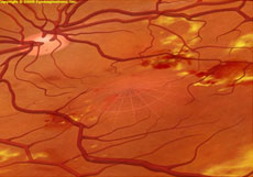
Early DiabeticRetinopathy
NPDR (Non-proliferative)
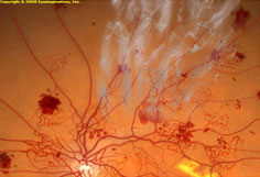
Advanced DiabeticRetinopathy
PDR (Proliferative)
This type of diabetic retinopathy is called nonproliferative diabetic retinopathy (NPDR). It’s called that because at this point, new blood vessels aren’t growing (proliferating). NPDR can be described as mild, moderate or severe. When you have NPDR, the walls of the blood vessels in your retina weaken. Tiny bulges called microaneurysms protrude from the vessel walls, sometimes leaking or oozing fluid and blood into the retina. As the condition progresses, the smaller vessels may close and the larger retinal vessels may begin to dilate and become irregular in diameter. Nerve fibers in the retina may begin to swell. Sometimes the central part of the retina (the macula) begins to swell, too. This is known as macular edema.
Proliferative diabetic retinopathy (PDR) is the most severe type of diabetic retinopathy. It’s called proliferative because at this stage, new blood vessels begin to grow in the retina. These new blood vessels are abnormal. They may grow or leak into the clear, jelly-like substance that fills the center of your eye (vitreous). Eventually, scar tissue stimulated by the growth of new blood vessels may cause the retina to detach from the back of your eye. If the new blood vessels interfere with the normal flow of fluid out of the eye, pressure may build up in the eyeball, causing glaucoma. This can damage the nerve that carries images from your eye to your brain (optic nerve).
Complications
Complications can lead to serious vision problems including:
Tests and Diagnosis
Diabetic retinopathy is best diagnosed with a dilated eye exam. For this exam, your eye doctor will place drops in your eyes that make your pupils open widely. This allows your doctor to get a better view inside your eye. The drops may cause your close vision to be blurry until they wear off several hours later.
During the exam, your eye doctor will look for:
- Presence or absence of a cataract
- Abnormal blood vessels
- Swelling, blood or fatty deposits in the retina
- Growth of new blood vessels and scar tissue
- Bleeding in the clear, jelly-like substance that fills the center of the eye (vitreous)
- Retinal detachment
- Abnormalities in your optic nerve
In addition, your eye doctor may:
- Test your vision
- Measure your eye pressure to test for glaucoma.
Fluorescein angiography
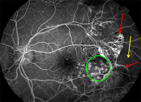
Fluorescein angiography
As part of the eye exam, your doctor may do a retinal photography test called fluorescein angiography. First, your doctor will dilate your pupils and take pictures of the inside of your eyes. Then your doctor will inject a special dye into your arm. More pictures will be taken as the dye circulates through your eyes. Your doctor can use the images to pinpoint blood vessels that are closed, broken down or leaking fluid.
Optical coherence tomography
Your eye doctor may request an optical coherence tomography (OCT) exam. This imaging test provides cross-sectional images of the retina that show the thickness of the retina, which will help determine whether fluid has leaked into retinal tissue. Later, OCT exams can be used to monitor how treatment is working.
Treatment and Drugs
Treatment depends largely on the type of diabetic retinopathy you have. Your treatment will also be affected by how severe your retinopathy is, and how it has responded to previous treatments.
Early diabetic retinopathy
If you have nonproliferative diabetic retinopathy (NPDR), you may not need treatment right away. However, your eye doctor will closely monitor your eyes to determine if you need treatment.
It may also be helpful to work with your diabetes doctor (endocrinologist) to find out if there are any additional steps you can take to improve your diabetes management. The good news is that when diabetic retinopathy is in the mild or moderate stage, good blood sugar control can usually slow the progression of diabetic retinopathy.
Advanced diabetic retinopathy
If you have proliferative diabetic retinopathy, you’ll need prompt surgical treatment. Sometimes surgery is also recommended for severe nonproliferative diabetic retinopathy. Depending on the specific problems with your retina, options may include:
Surgery often slows or stops the progression of diabetic retinopathy, but it’s not a cure. Because diabetes is a lifelong condition, future retinal damage and loss of vision are possible. Even after treatment for diabetic retinopathy, you’ll need regular eye exams. At some point, additional treatment may be recommended.
Hear from our patients!
Treating Families - Not Just Their Conditions